

Magnetic Resonance Angiography (MRA)
What to Expect
On Arrival
During the Procedure
After the Procedure
Frequently Asked Questions
Magnetic Resonance Imaging (MRI)
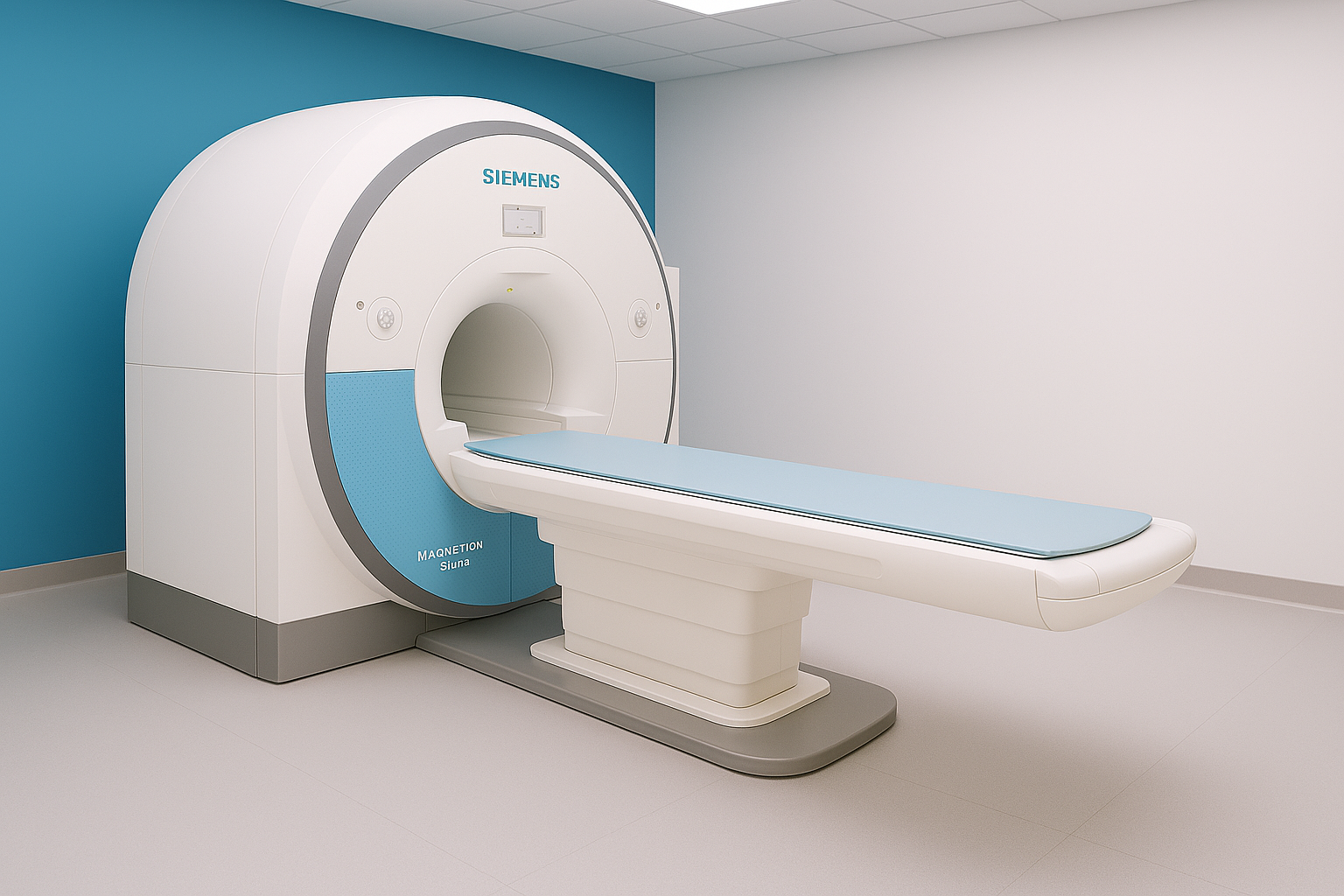
MRI is a medical imaging exam that uses nuclear magnetic resonance to visualize internal structures of the body in detail. There is no radiation. MRI’s help Radiologist examine organs, muscles, joints,ligaments, muscles, bones, brain, spine and soft tissue anatomy. An MRI creates two-dimensional or three-dimensional images of any part of the body. MRIs provide good visual contrast between the fat and water content of the body which makes them especially useful in revealing abnormalities like tumors, inflammation and infection.
Mammography
Mammography is a specialized medical imaging exam that uses low-dose X-rays to create detailed images of breast tissue. It is mainly used to screen for and detect breast cancer early, often before a lump can be felt. Mammograms can help doctors identify masses, calcifications, or other changes in breast tissue and are an important tool for early detection and monitoring of breast health. Modern digital and 3D mammography technology improves image clarity and detection while keeping radiation exposure low and patient comfort a priority.