

Ultrasound
Ultrasound is a simple, comfortable imaging test that uses sound waves to create live pictures of what is happening inside your body. There is no radiation; just clear, helpful images that give your provider the information they need. Whether you are checking an organ, looking at blood flow, or monitoring a pregnancy, we are here to make the experience easy and relaxing from start to finish.
What to Expect
On Arrival
You will be greeted and checked in by one of our friendly staff at the front‑desk.
Once you are ready, we will walk you to the ultrasound room. Depending on the type of exam, you may change into a gown, but we will guide you through it so you know exactly what to do.
Our sonographer will start by walking you through the exam so you know exactly what to expect. They may ask you a few simple questions about your symptoms or medical history. This helps us know what to focus on in real-time while we follow the appropriate exam protocol.
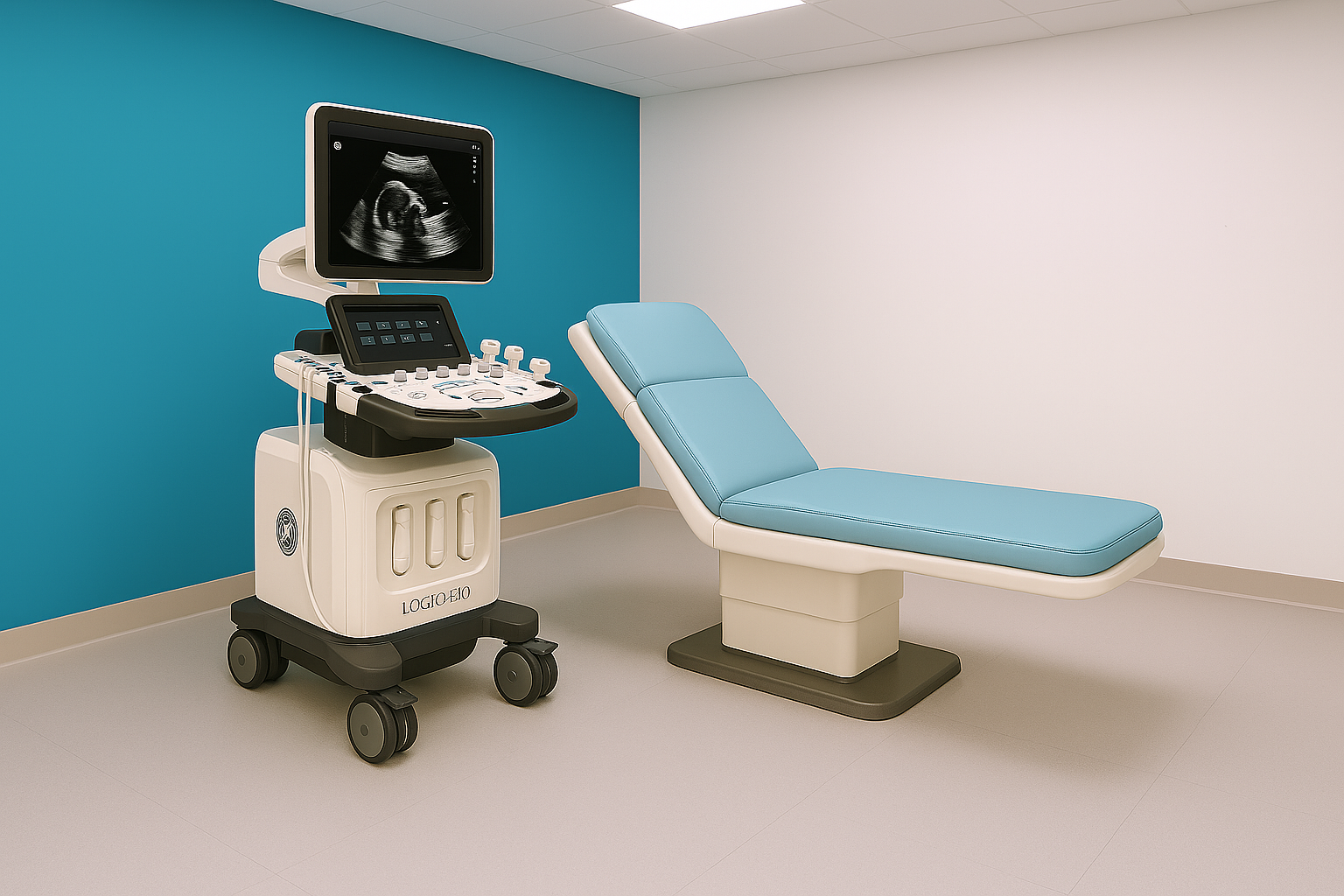
During the Procedure
You will lie comfortably on the exam table, and we will apply a warm, water‑based gel to the area being imaged. The gel helps the sound waves travel clearly so we can capture the best possible images.
The sonographer will use a small handheld device called a transducer to gently scan the area. You may be asked to shift your position or briefly hold your breath so we can capture the clearest images.
After the Procedure
When you are done, we will give you towels so you can wipe off the gel. You can then get dressed.
There is no recovery time. You may resume normal activities immediately after the ultrasound.
Frequently Asked Questions
Dual-energy X-ray Absorptiometry (DEXA or DXA)
Dual-energy X-ray Absorptiometry (DEXA or DXA) is a specialized imaging exam that uses very low-dose X-rays to measure bone density and body composition. It is used to diagnose osteoporosis, assess fracture risk, and detect early bone loss. DEXA scans also provide detailed measurements of fat and lean muscle distribution, supporting fitness, nutrition, and wellness goals.
X-ray (Radiography)