

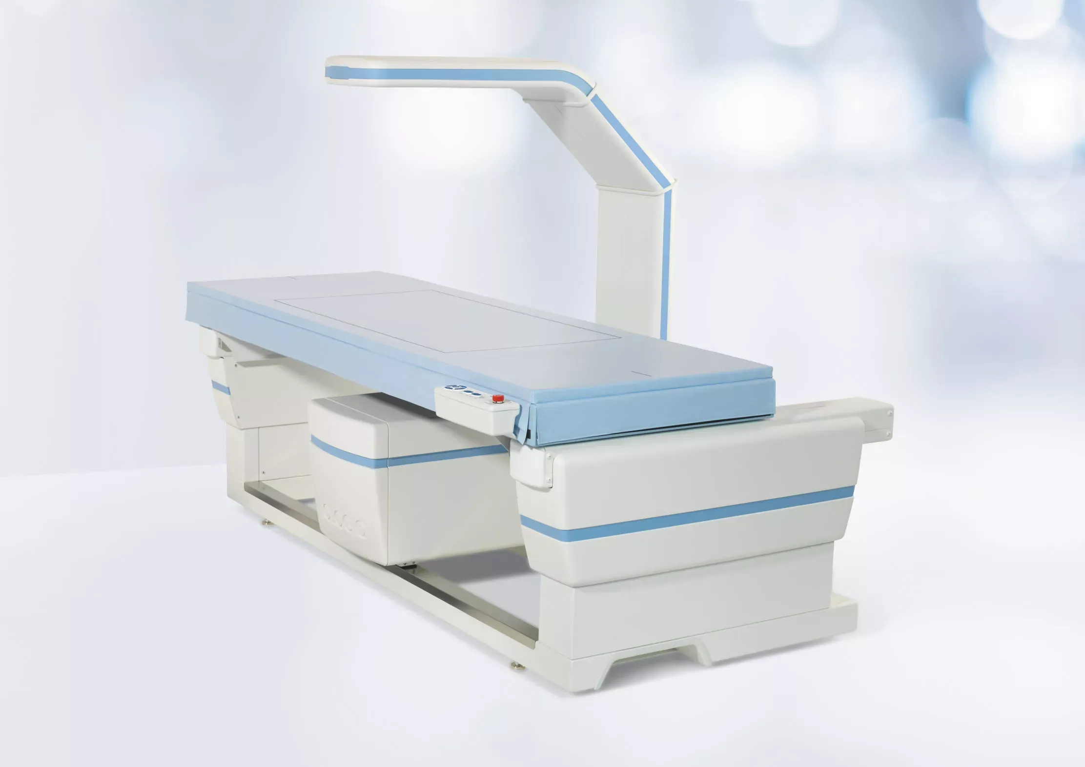
Dual-energy X-ray Absorptiometry (DEXA or DXA)
What to Expect
On Arrival
During the Procedure
After the Procedure
Frequently Asked Questions
Mammography
Mammography is a specialized medical imaging exam that uses low-dose X-rays to create detailed images of breast tissue. It is mainly used to screen for and detect breast cancer early, often before a lump can be felt. Mammograms can help doctors identify masses, calcifications, or other changes in breast tissue and are an important tool for early detection and monitoring of breast health. Modern digital and 3D mammography technology improves image clarity and detection while keeping radiation exposure low and patient comfort a priority.
Ultrasound
Ultrasound is a simple, comfortable imaging test that uses sound waves to create live pictures of what is happening inside your body. There is no radiation; just clear, helpful images that give your provider the information they need. Whether you are checking an organ, looking at blood flow, or monitoring a pregnancy, we are here to make the experience easy and relaxing from start to finish.